Innovic Medical Bacterial Cell Structure
A differentiated resource for producing a labelled diagram of a bacterium. There are two main versions of this activity, but both of them require the information cards. These can be printed out and placed around the room.
Bacterial Cell Structure and Function
bacteria, any of a group of microscopic single-celled organisms that live in enormous numbers in almost every environment on Earth, from deep-sea vents to deep below Earth's surface to the digestive tracts of humans. Bacteria lack a membrane-bound nucleus and other internal structures and are therefore ranked among the unicellular life-forms.
Labelled Diagram Of Bacteria
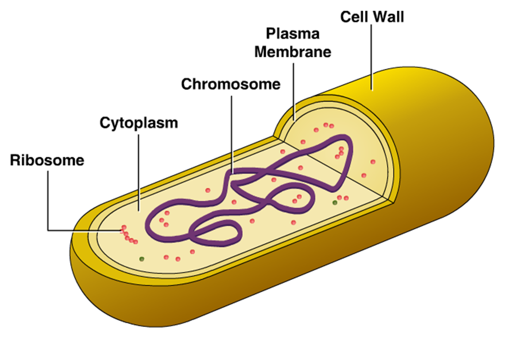
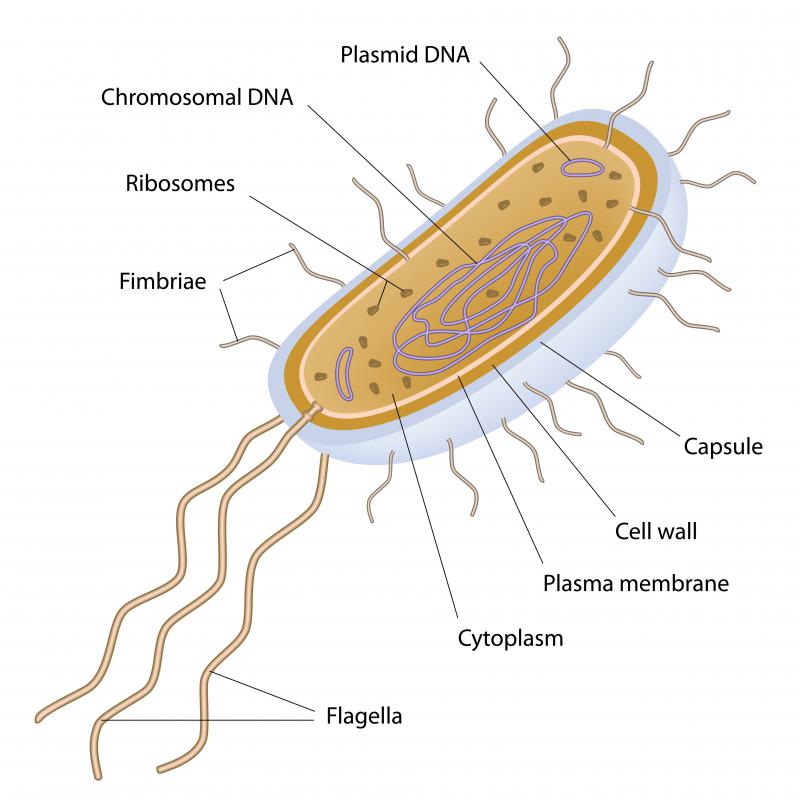
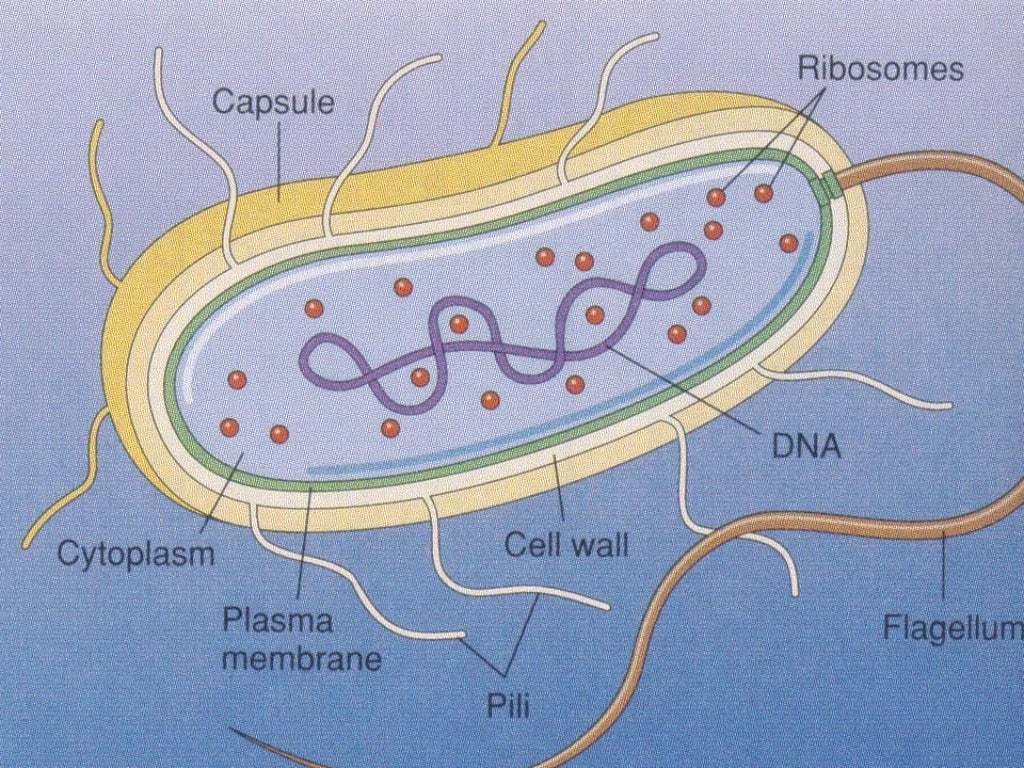
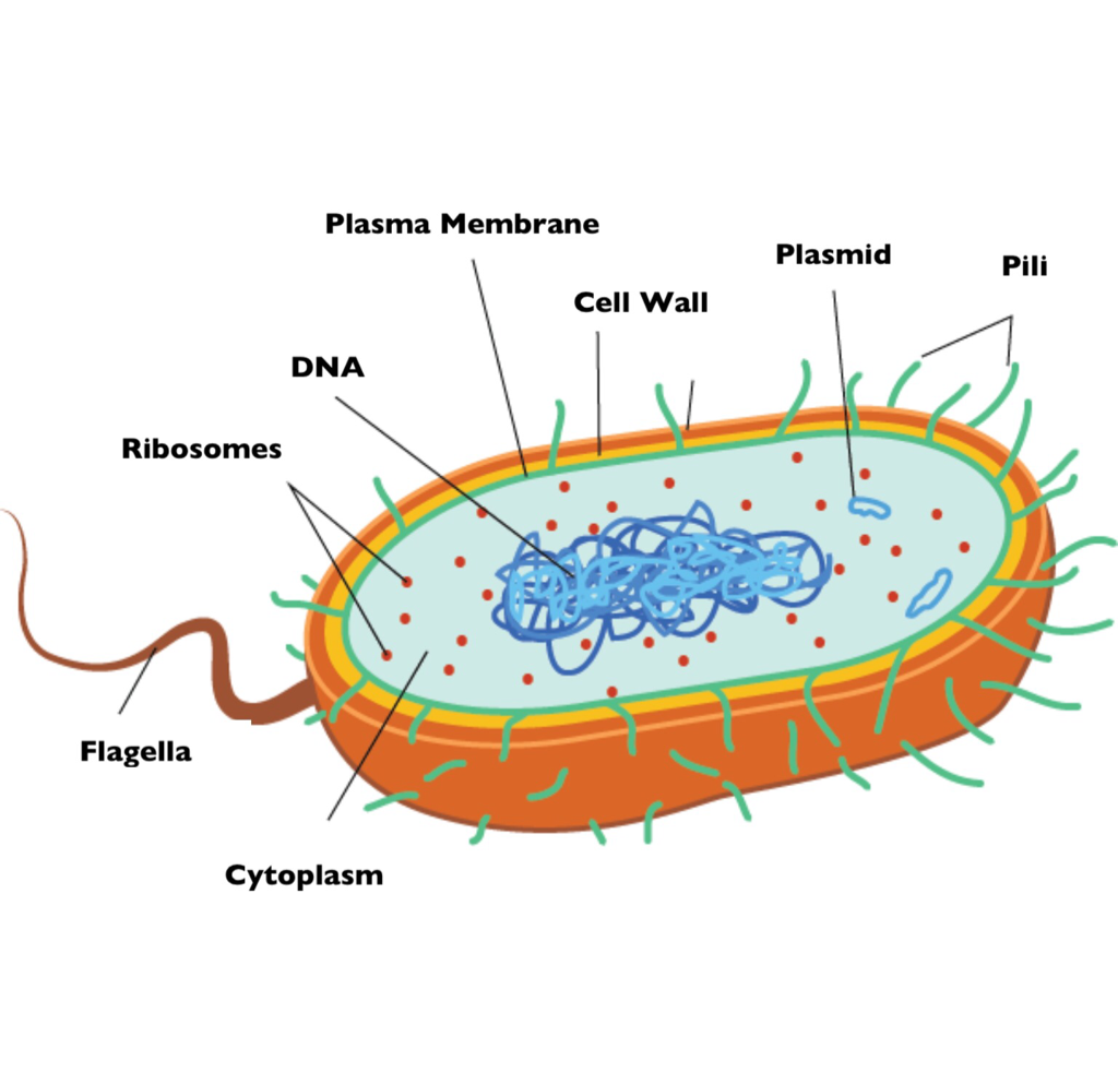
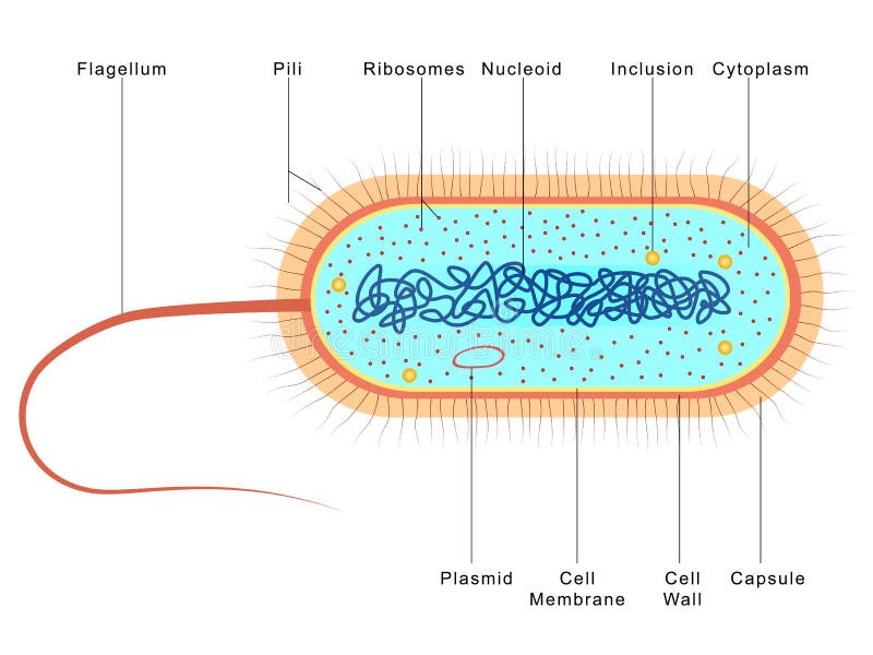
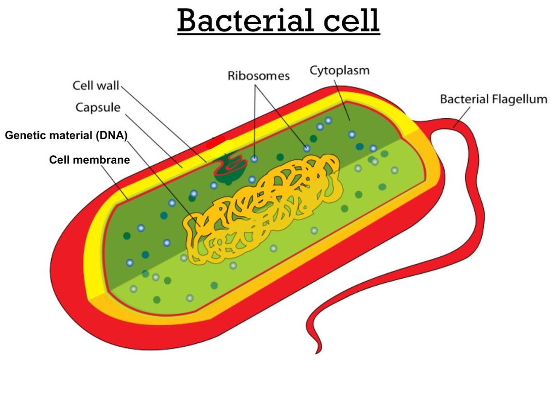
Bacteria Diagram The bacteria diagram given below represents the structure of a typical bacterial cell with its different parts. The cell wall, plasmid, cytoplasm and flagella are clearly marked in the diagram. Bacteria Diagram representing the Structure of Bacteria Ultrastructure of a Bacteria Cell
Bacterial Structure Plantlet
The bacteria shapes, structure, and labeled diagrams are discussed below. Table of Contents [ show] Sizes The sizes of bacteria cells that can infect human beings range from 0.1 to 10 micrometers. Some larger types of bacteria such as the rickettsias, mycoplasmas, and chlamydias have similar sizes as the largest types of viruses, the poxviruses.
Bacteria Cell Vector Art, Icons, and Graphics for Free Download
These can rotate or move in a whip-like motion to move the bacterium. Plant and bacterial cell walls provide structure and protection. Only plant cell walls are made from cellulose. The DNA of.
What is a Bacterium? (with pictures)
Biology teaches use that bacteria tend to be unicellular organisms with a peculiar structure. Featuring in this page is an interactive bacteria labelled diagram. It features an annotated diagram with labels to drag and drop at the correct position. This worksheet teaches students the structure of bacteria in a fun way.

Bacterium Cell Labeled
It also means that you—for some definition of the word you—actually consist of both of the major types of cells: prokaryotic and eukaryotic. All cells fall into one of these two broad categories. Only the single-celled organisms of the domains Bacteria and Archaea are classified as prokaryotes— pro means before and kary means nucleus.
Bacterial Cell Color Diagram of Organelles Inside the Cell Wall for Science and Biology Concepts
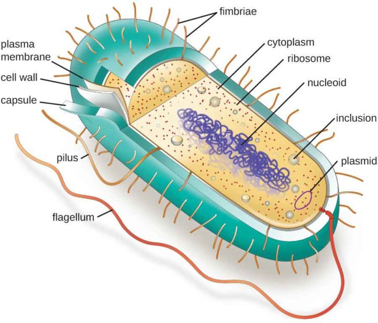
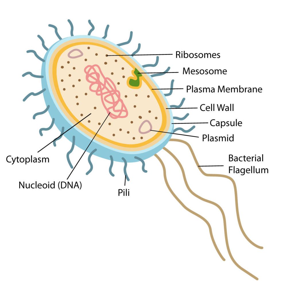
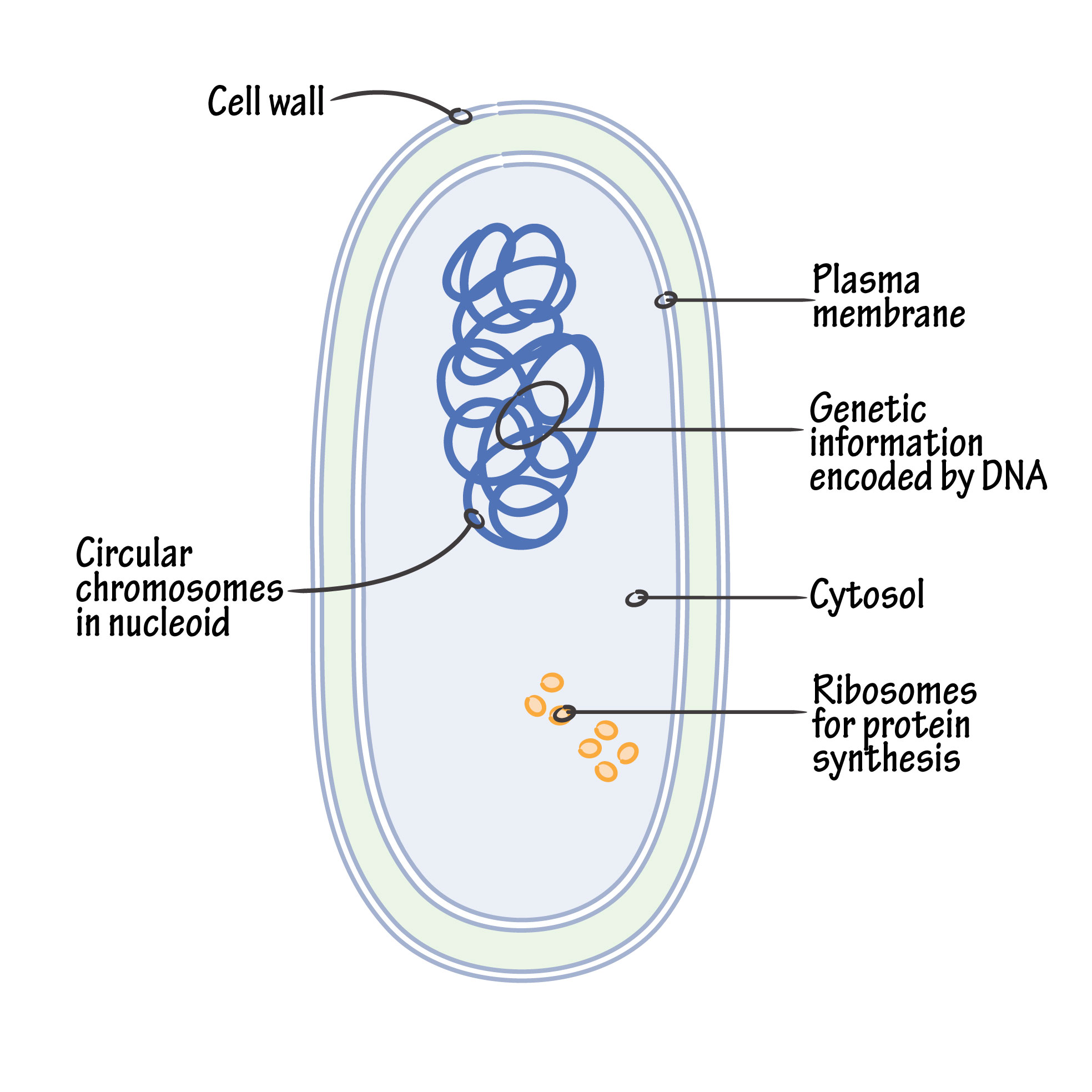
Key points: Prokaryotes are single-celled organisms belonging to the domains Bacteria and Archaea. Prokaryotic cells are much smaller than eukaryotic cells, have no nucleus, and lack organelles. All prokaryotic cells are encased by a cell wall. Many also have a capsule or slime layer made of polysaccharide.
Bacteria Diagram Photograph by Monica Schroeder
The below mentioned article provides a diagram of bacteria along with additional information as follows:- 1. Occurrence and Distribution of Bacteria 2. Size of Bacteria 3. Forms 4. Staining Bacteria (Gram Reaction). Occurrence and Distribution of Bacteria: The bacteria constitute a highly specialised group of one-celled plants.
Bacterial cell anatomy in flat style. Vector modern illustration. Labeling structures on a
Cell wall The structure of peptidoglycan. The cell envelope is composed of the cell membrane and the cell wall.As in other organisms, the bacterial cell wall provides structural integrity to the cell. In prokaryotes, the primary function of the cell wall is to protect the cell from internal turgor pressure caused by the much higher concentrations of proteins, and other molecules inside the.
Bacterial structure and morphology by Dr. Shireen Rafiq (RMC)
Diagram showing the relative sizes of some very small things including bacteria, which are typically around 1 to 2 μm in diameter (Source: Michigan Nanotechnology Institute for Medicine and Biological Sciences ). Image - Text Version Even though they are small, bacterial cells have many different parts.
Bacterial Structure Plantlet
Structure and Function of a Typical Bacterial Cell with Diagram Animesh Sahoo August 14, 2021 Bacteria are unicellular. Their structure is a very simple type. Bacteria are prokaryotes because they do not have a well-formed nucleus. A typical bacterial cell is structurally very similar to a plant cell.
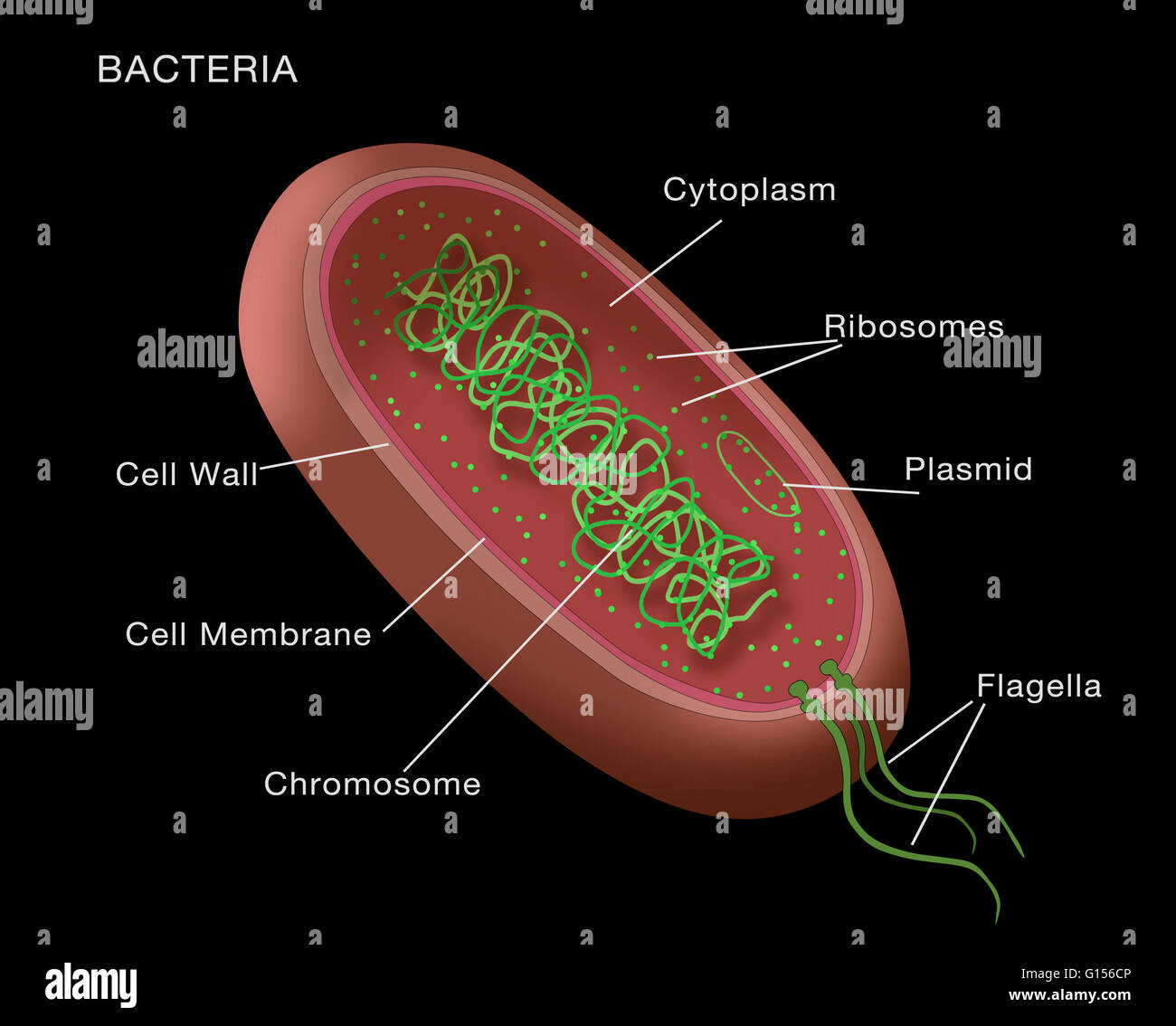
Illustration of a typical bacterium, with key parts (cell membrane, cytoplasm, flagella, etc
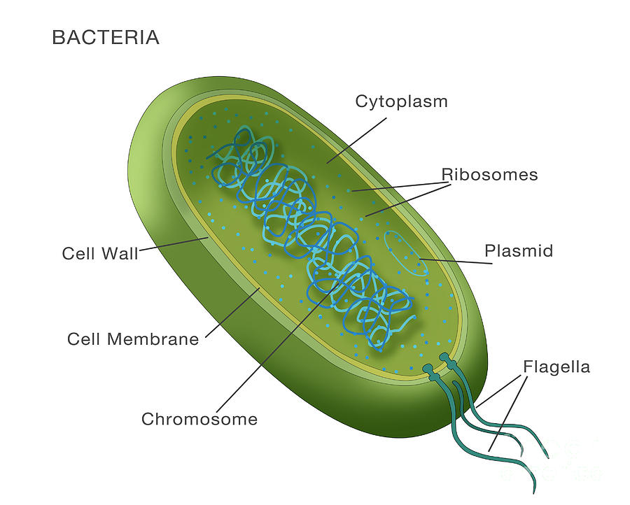
Bacteria (sing. bacterium) are unicellular prokaryotic microorganisms which divide by binary fission. They do not possess nuclear membrane and the nucleus consists of a single chromosome of circular double-stranded DNA helix (Fig. 1.1). Flagella: ADVERTISEMENTS:
Bacteria Grade 11 Biology Study Guide
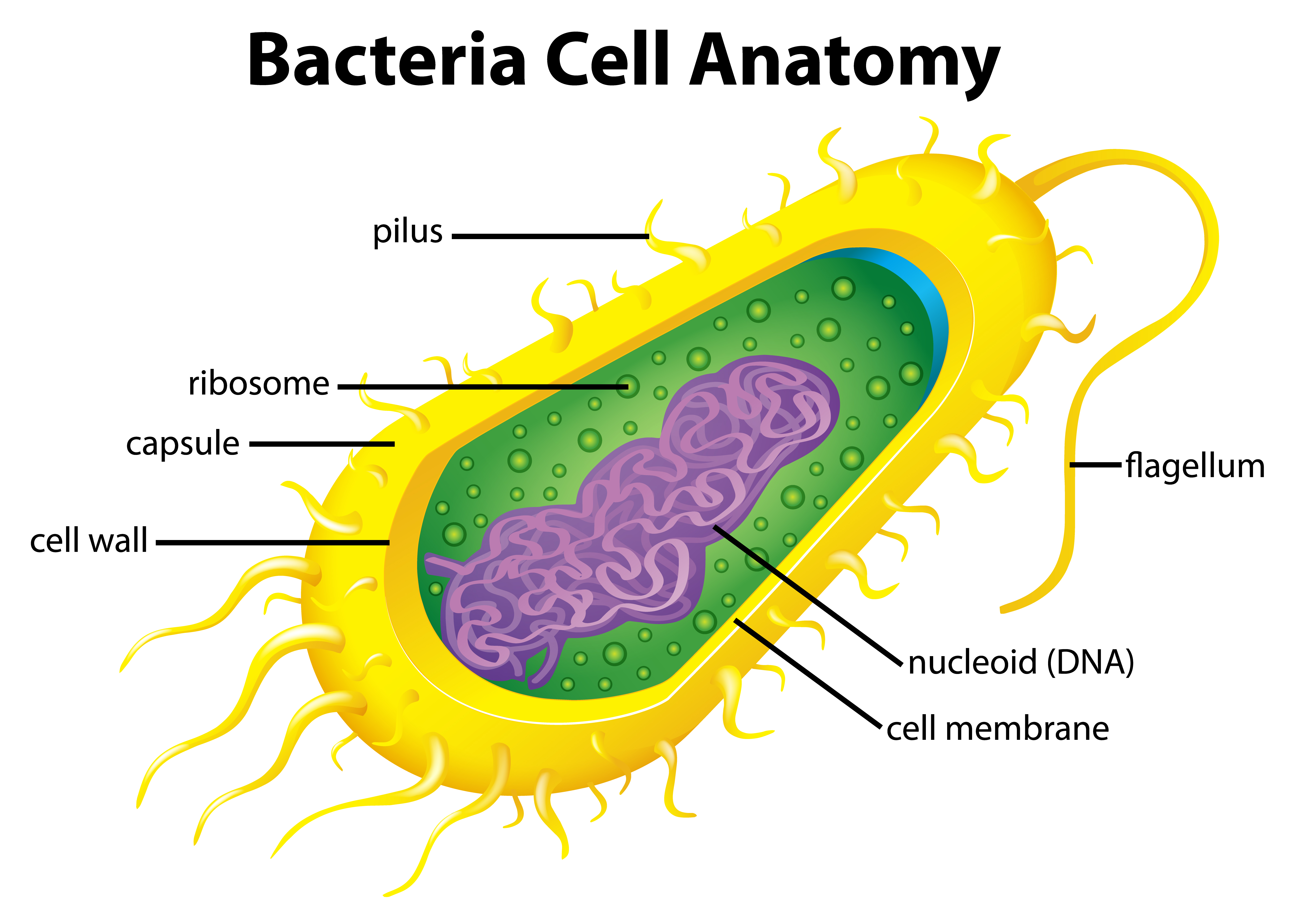
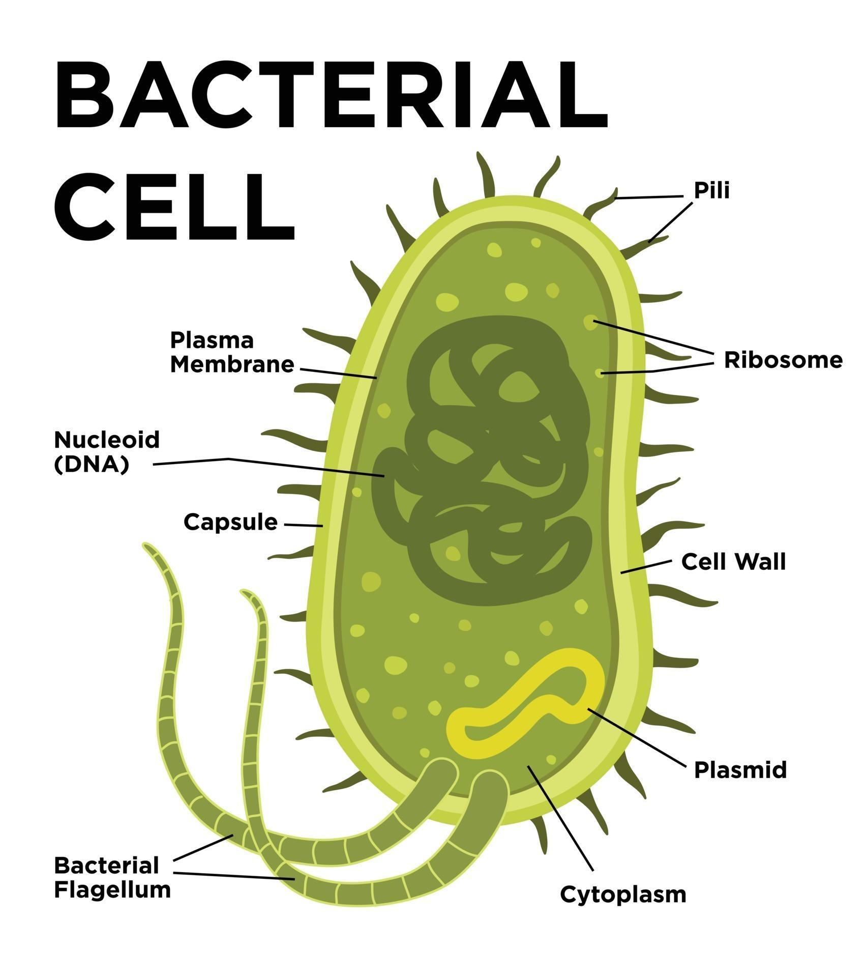
Bacteria Diagram with Labels Bacterial cells have simpler internal structures like Pilus (plural Pili), Cytoplasm, Ribosomes, Capsule, Cell Wall, Plasma membrane, Plasmid, Nucleoid, Flagellum, etc. Labeled Bacteria diagram Eukaryotes have been shown to be more recently evolved than prokaryotic microorganisms.
Anatomy of Bacteria stock vector. Illustration of labelled 43965779
A detailed bacteria labelled diagram for your classroom Bacteria in the classroom - definitely not something we really like to think about! But, it's an essential consideration, especially when children are learning about different cells and organisms.
Bacteria Ms A Science Online
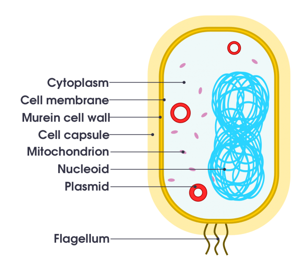
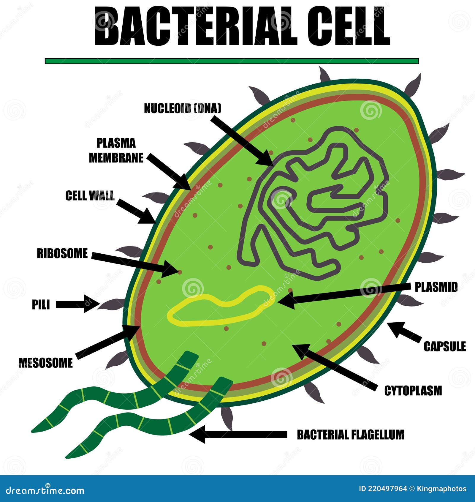
In this article we will discuss about the cell structure of bacteria with the help of diagrams. A bacterial cell (Fig. 2.5) shows a typical prokaryotic structure. The cytoplasm is enclosed by three layers, the outermost slime or capsule, the middle cell wall and inner cell membrane. The major cytoplasmic contents are nucleoid, plasmid, ribosome.